EEGBCI Technology
ECoG and Functional Mapping: Christoph Guger
More than 3,500 participants from all over the world met at the BCI & Neurotechnology Spring School. Stretching from Argentina to Japan and New Zealand, this global community made up the largest BCI event in history. We report the latest advances in invasive BCI, functional brain mapping and brain stimulation from day 3.
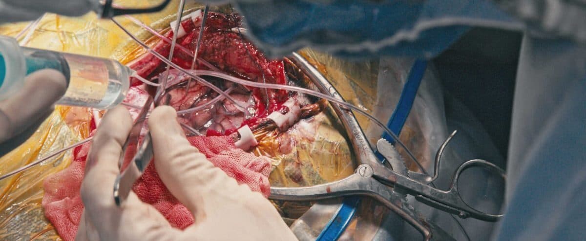
ECoG and Functional Mapping: Christoph Guger
As on every day, the first speaker of the day was Christoph Guger. Today Christoph covered ECoG (electrocorticography) and functional mapping.
Christoph summed up the difference in spatial resolution between EEG and ECoG data as follows:
- What EEG makes possible
- It is possible to distinguish between left-hand and right-hand motor imagery, but it is not possible to tell individual finger movements apart.
- What ECoG makes possible
- When electrodes are implanted directly on the cortex, individual finger movements can be clearly distinguished using 80–200 Hz high-gamma activity. This makes it possible to identify the cortical areas responsible for each finger movement.
Thanks to this superior spatial resolution, g.tec developed a high-gamma mapping tool called cortiQ. With this system, neurosurgeons can map a person’s most important, indispensable brain functions within just a few minutes.
This work requires close collaboration with neurosurgeons such as Anthony Ritaccio and Kyousuke Kamada. The system provides a mapping paradigm that makes it possible to identify the cortical areas responsible for the hands, lips, eyes, tongue, mathematical centres, face areas, colour and black-and-white decoding areas, and many more.
This information is critical for the neurosurgeon to know which tissue must not be touched during surgery.
Where Is the cortiQ System Used?
- In intensive-care monitoring of epilepsy patients
- In identifying the “eloquent cortex” during surgery on tumour patients
How to Set Up ECoG BCI Experiments — Fan Cao
Fan Cao explained how ECoG BCI experiments are run. Depending on the surgical requirements, ECoG grids are placed over the sensorimotor area, the temporal lobe, the visual cortex and/or other areas. This planning is done with cortiQ’s Montage Creator.
Next, an experimental paradigm is created that defines the tasks the patient has to perform. Both of these steps are usually completed during surgical planning.
When the patient enters brain mapping, cortiQ replays the experimental paradigm so that the patient performs the task. The whole mapping procedure usually takes 3–5 minutes.
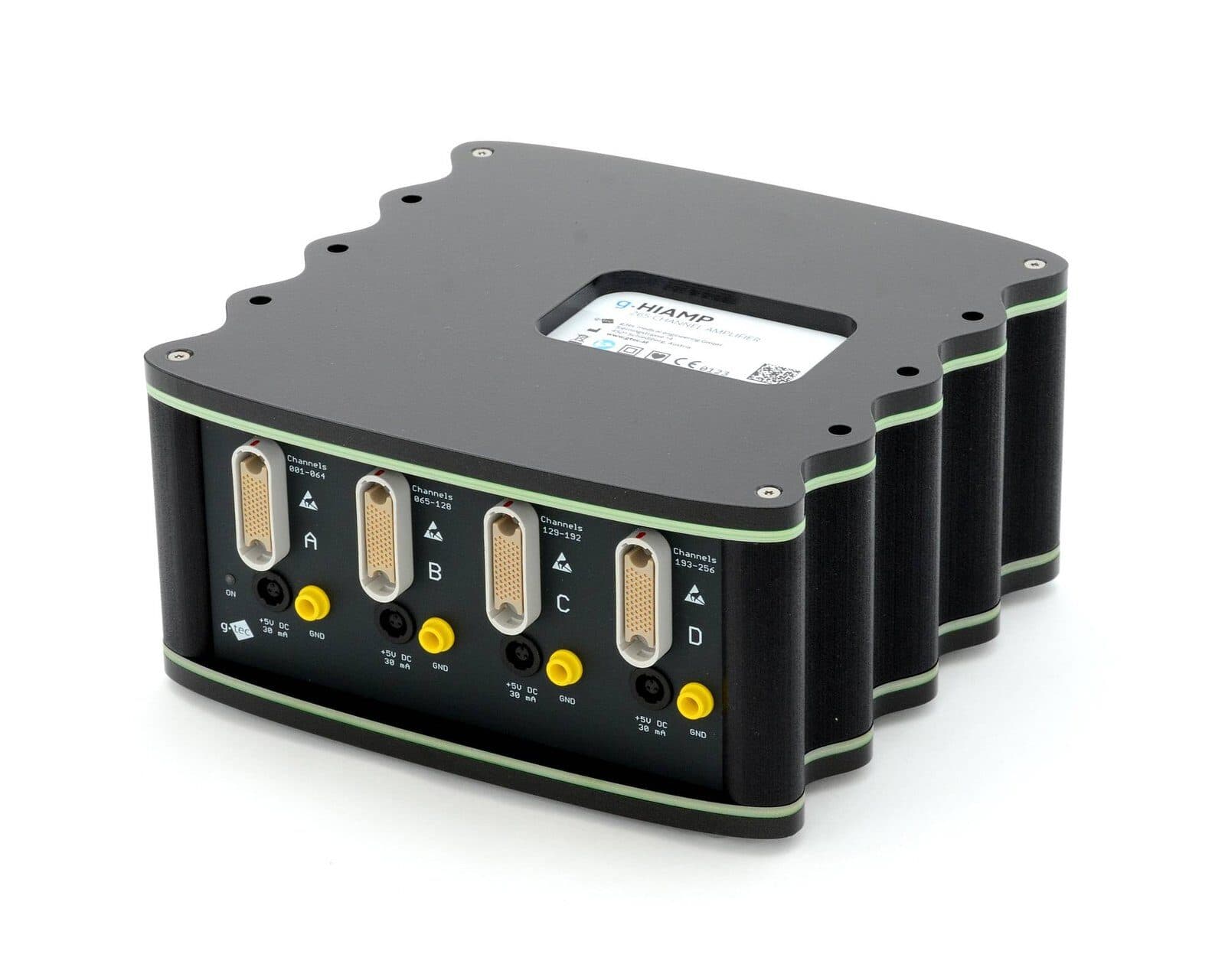
Fan also showed how to configure a complete closed-loop experiment with ECoG data recorded with g.HIamp, using the g.HIsys rapid prototyping software environment. He showed the audience how a real-time system can recognise 16 different types of visual stimuli presented to the patient. One practical application of this example is decoding in real time whether the patient is seeing a face or a symbol on a computer screen.
Optimising Open- and Closed-Loop Brain Stimulation — Johannes Grünwald
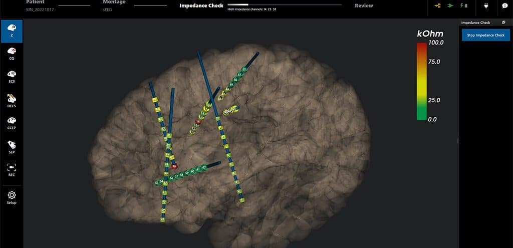
Johannes Grünwald presented the critical parameters to watch when performing electrical stimulation with surface ECoG electrodes and with the depth electrodes used for DBS (deep brain stimulation).
The g.HIsys software environment can be used to design real-time experiments. It allows neural spikes and ECoG data to be recorded with biosignal amplifiers.
The new g.Estim Switching Unit is controlled in real time, allowing the stimulation site to be selected on the fly. It disconnects the amplifier during stimulation, guaranteeing that all of the current is applied to the cortex.
What Do Invasive BCIs Offer Us? — Milena Korostenskaja
Milena Korostenskaja of the Neuroapproaches Institute discussed in detail how ECoG, MEG and fMRI recordings are carried out in hospitals and which technologies they require.
Only a few weeks earlier, Milena and her team had operated on a newborn baby to treat epilepsy. Treating epilepsy in young children is especially critical: without intervention, more and more neurons are affected.
Using grid, strip and depth electrodes in her ECoG studies, Milena described how high-gamma mapping technology is validated against electrical current stimulation, MEG and fMRI. Her research clearly showed that more information can be obtained with high-gamma mapping and that the method is more sensitive than electrical current stimulation.
Closed-Loop Neuromodulation for Parkinson’s — Nuri Firat Ince
Nuri Firat Ince of the University of Houston’s Department of Biomedical Engineering discussed deep brain stimulation (DBS) techniques for Parkinson’s patients.
Implanted electrodes can stop these patients’ tremors with electrical stimulation. Beta oscillations in the LFP decode the pathological state in these patients and guide systems that apply deep brain stimulation to the STN region of the thalamus when needed.
Nuri also presented the development of biomarkers that indicate different types of Parkinson’s. In some Parkinson’s patients tremor dominates, in others walking difficulty. For these patients, phase-amplitude coupling can be used as a biomarker. His team is currently making chronic recordings in Parkinson’s patients to investigate whether and how the treatment helps.
Reconstructing Speech with ECoG BCI — Dean Krusienski
Dean Krusienski of Virginia Commonwealth University gave a comprehensive overview of the groups working on speech production and reconstruction: Eddie Chang and Bob Knight at Berkeley and Nima Mesgarani at Columbia University are among the pioneers in the field.
Working with Northwestern University, Dean records ECoG data in intraoperative studies. His team uses neural networks to reconstruct the speech signal efficiently. As new data arrive, they are compared with the training data to build a lookup table, and speech is synthesised from it.
The log power of the gamma band between 70 and 170 Hz is used as the input feature. This approach makes it possible to reconstruct words such as “shook”, “steer” and “pave” with high accuracy and to understand the resulting signal.
Key takeaway: this research shows that brain–computer interface technology can reconstruct not only movement but the human voice and language itself. For patients who are paralysed or have lost the ability to speak, this opens a door of hope.